Before you can understand any organ, drug or disease, you need to understand the cell. Almost every medicine you will study works by changing something inside or on the surface of a cell. This post explains the cell and each of its parts in simple language.
What is a cell?
The cell is the smallest unit of life. The simplest living things, such as bacteria, are made of just one cell. Higher animals, including humans, are made of trillions of cells working together, like a huge society where every member has a job.
In the body there are brain cells (neurons), skin cells, liver cells, stomach cells, muscle cells, blood cells and many more. Each type looks different and does a different job. For example:
- Nerve cells are long and thin so they can carry messages over long distances.
- Red blood cells are small, flexible discs with no nucleus, so they can squeeze through tiny vessels and carry more oxygen.
- Muscle cells are packed with proteins that can shorten and pull.
Even though they look different, almost all human cells share the same basic parts.
Animal cell vs. plant cell
Human cells are animal cells. They are eukaryotic, which means their DNA is kept inside a proper nucleus with a membrane around it, and they contain many membrane-bound organelles. Unlike plant and fungal cells, animal cells do not have a cell wall. They are covered only by a thin, flexible plasma membrane. They also do not have chloroplasts, because animals do not make their own food from sunlight.
How big is a cell?
Most human cells are between 5 and 50 micrometres wide (1 micrometre = one thousandth of a millimetre). That is far too small to see without a microscope. Some cells are exceptionally large; the mature egg cell (ovum) is about 120 micrometres across and is just visible to the naked eye. Nerve cells can be over a metre long, although they are very thin.
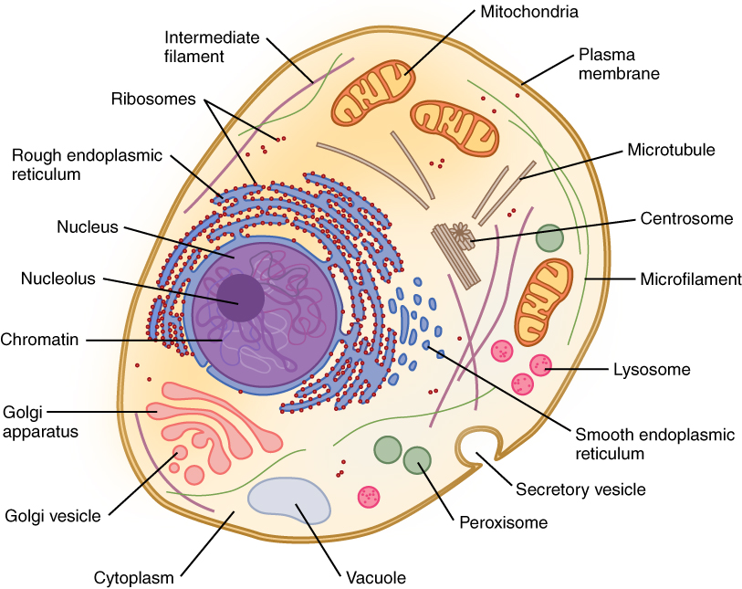
The three main regions
- Plasma membrane: the outer boundary.
- Cytoplasm: everything between the membrane and the nucleus. It includes a jelly-like fluid called cytosol and the organelles floating in it.
- Nucleus: the control centre containing DNA.
A simple way to remember the organelles is to imagine the cell as a factory. Each part below has a factory job.
Parts of the cell explained
1. Plasma (cell) membrane: the gate and security guard
All living cells have a plasma membrane around their contents. It is made of a double layer of fat molecules (the phospholipid bilayer) with proteins floating in it. In bacteria, the membrane sits inside a rigid cell wall, but animal cells have only the membrane to hold and protect their contents.
The membrane is selectively permeable: it lets some substances in and keeps others out. Oxygen and carbon dioxide pass through easily, while glucose and ions need special protein channels or carriers. Many drug targets, called receptors, sit on this membrane.
2. Nucleus: the head office
The nucleus is the largest organelle and the information centre of the cell. It has two main jobs:
- It stores the hereditary material (DNA), organised into chromosomes. Humans have 46 chromosomes in most cells.
- It controls the cell's activities, including growth, metabolism, protein making and cell division.
It is surrounded by a double membrane called the nuclear envelope, which has small holes (nuclear pores) so that messages (RNA) can leave. Inside is a dark spot called the nucleolus, where ribosomes are made.
3. Ribosomes: the workers
Ribosomes are tiny structures made of about 60% RNA and 40% protein. Their job is to make proteins by reading the instructions sent from the DNA. Some float freely in the cytoplasm (making proteins for use inside the cell); others are attached to the rough endoplasmic reticulum (making proteins to be exported). Human ribosomes are slightly different from bacterial ones, which is why some antibiotics can kill bacteria without harming us.
4. Endoplasmic reticulum (ER): the production line
The ER is a network of folded membranes and sacs that makes, processes and transports chemicals for use inside and outside the cell. It is connected to the nuclear envelope, acting like a pipeline between the nucleus and the cytoplasm. There are two types:
- Rough ER is covered with ribosomes and makes proteins.
- Smooth ER has no ribosomes. It makes lipids (fats and steroid hormones) and, in liver cells, helps break down drugs and toxins.
5. Golgi apparatus: packing and shipping department
The Golgi apparatus looks like a stack of flat pancakes. It receives proteins and fats from the ER, modifies, sorts and packs them into small bubbles called vesicles, and sends them to where they are needed, either inside the cell or outside it (secretion). Cells that secrete a lot, like pancreatic cells that release insulin, have a large Golgi.
6. Mitochondria: the power plant
Mitochondria are oblong organelles found in the cytoplasm of almost every cell. They are the powerhouses of the cell: they use oxygen and nutrients (like glucose and fat) to make ATP (adenosine triphosphate), the energy "currency" that powers nearly every activity in the cell. This process is called cellular respiration.
Cells that need a lot of energy, such as heart muscle cells, have thousands of mitochondria. Mature red blood cells have none, so they make their energy from glucose without using oxygen. Interestingly, mitochondria have their own small piece of DNA, inherited only from the mother.
7. Lysosomes: the recycling and waste-disposal unit
Lysosomes are small sacs filled with powerful digestive enzymes. They break down worn-out cell parts, waste, and germs or debris taken in from outside the cell into simple compounds, which the cell can reuse as building materials. If a lysosome bursts, its enzymes can digest the whole cell, which is why lysosomes are sometimes called "suicide bags".
8. Peroxisomes: the cleaners
Peroxisomes are small, round organelles bound by a single membrane. They are the most common type of microbody. They break down fatty acids and neutralise toxic substances such as hydrogen peroxide (using the enzyme catalase). Liver and kidney cells have many peroxisomes because they do a lot of detoxifying.
9. Centrioles: the organisers of cell division
Centrioles are a pair of small cylinders found near the nucleus in animal cells. Each one is made of nine triplets of microtubules (a "9 x 3" pattern). They are self-replicating and help organise the spindle that pulls chromosomes apart during cell division, although cells can still divide without them.
10. Cytoskeleton: the steel frame
The cytoskeleton is a network of protein fibres that gives the cell its shape, holds organelles in place and helps the cell move. It has three types of fibres:
| Fibre | Made of | Main job |
|---|---|---|
| Microfilaments (thinnest) | Actin | Shape and movement; in muscle they slide against myosin to cause contraction |
| Intermediate filaments | Various fibrous proteins (e.g. keratin) | Strong "ropes" that bear tension and keep the cell firm |
| Microtubules (thickest) | Tubulin | Tracks for transport inside the cell; form centrioles, cilia, flagella and the spindle in cell division |
Thick filaments are made of the protein myosin. In muscle cells, myosin pulls on actin and the muscle shortens.
11. Cilia and flagella: brushes and tails
These are hair-like projections from the cell surface, built from microtubules. For single-celled organisms they are needed to swim. In the human body:
- Cilia are short and many. They beat together to move fluid past the cell, for example sweeping mucus and dust up out of the airways, or moving the egg along the fallopian tube.
- Flagella are long and usually single. The only human cell with a flagellum is the sperm cell, which uses it to swim.
12. Endosomes, endocytosis and exocytosis: how big things get in and out
Some materials are too big to pass through the membrane. The cell handles them in two ways:
- Endocytosis (bringing in): the membrane folds inward (invaginates), wraps around the material and pinches off to form a bubble inside the cell called an endosome. When the cell "eats" solid particles such as bacteria, it is called phagocytosis; when it "drinks" fluid, it is called pinocytosis.
- Exocytosis (sending out): the reverse. A vesicle joins the membrane and releases its contents outside, for example when nerve cells release neurotransmitters or gland cells release hormones.
Quick summary table
| Organelle | Factory job | Function |
|---|---|---|
| Nucleus | Head office | Stores DNA, controls the cell |
| Plasma membrane | Gate | Controls what enters and leaves |
| Mitochondria | Power plant | Makes ATP (energy) |
| Ribosomes | Workers | Make proteins |
| Rough / smooth ER | Production line | Proteins / lipids and detox |
| Golgi apparatus | Packing and shipping | Modifies, packs and secretes |
| Lysosomes | Recycling | Digest waste and germs |
| Peroxisomes | Cleaners | Break down fatty acids and H2O2 |
| Centrioles | Organisers | Help in cell division |
| Cytoskeleton | Steel frame | Shape, support, movement |
- Cell = basic structural and functional unit of life.
- Human cells are eukaryotic animal cells: nucleus present, no cell wall, no chloroplasts.
- Most human cells: 5 to 50 micrometres. Largest: ovum (about 120 micrometres).
- Mitochondria = powerhouse (ATP). Ribosomes = protein synthesis. Lysosomes = "suicide bags".
- Rough ER = proteins; smooth ER = lipids and detoxification.
- Centrioles = 9 triplets of microtubules.
- Actin (thin filament) + myosin (thick filament) = muscle contraction.
- Mature red blood cells have no nucleus and no mitochondria.
- Ribosomes: antibiotics such as tetracyclines, macrolides (azithromycin) and aminoglycosides block bacterial ribosomes.
- Cell wall: penicillins and cephalosporins attack the bacterial cell wall, which human cells do not have. That is why they are relatively safe for us.
- Microtubules: cancer drugs like vincristine and paclitaxel stop cell division by acting on microtubules; colchicine (for gout) also binds tubulin.
- Smooth ER: liver enzymes here (cytochrome P450) break down many drugs, which explains many drug interactions.
- Membrane receptors: most drugs act by binding to receptors on the cell membrane.
No comments:
Post a Comment